Michael Hooker Microscopy Facility (MHMF.ORG)
![]()
|
|
Michael Hooker Microscopy Facility (MHMF.ORG) |
|
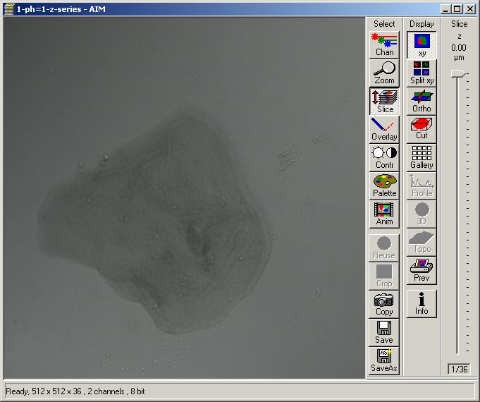
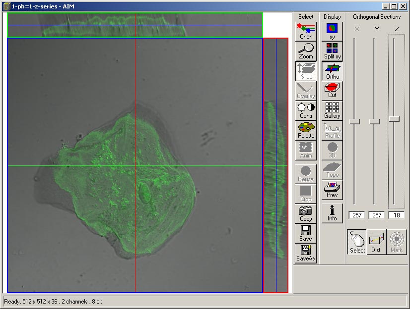
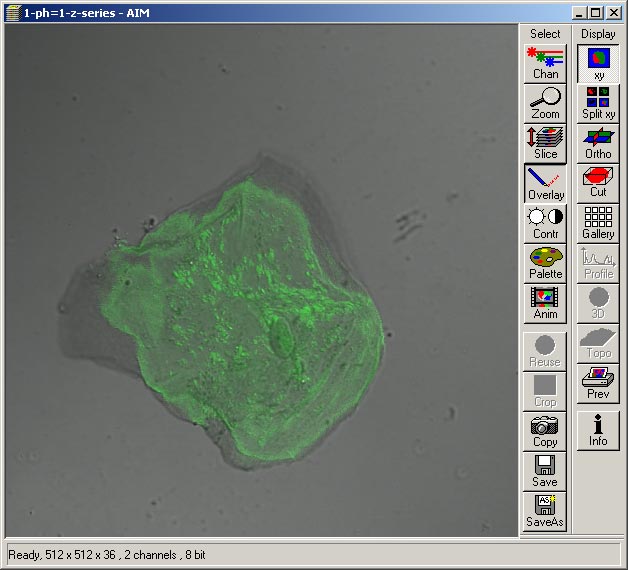
Viewing Zeiss 510 Confocal Image Databases
Introduction:
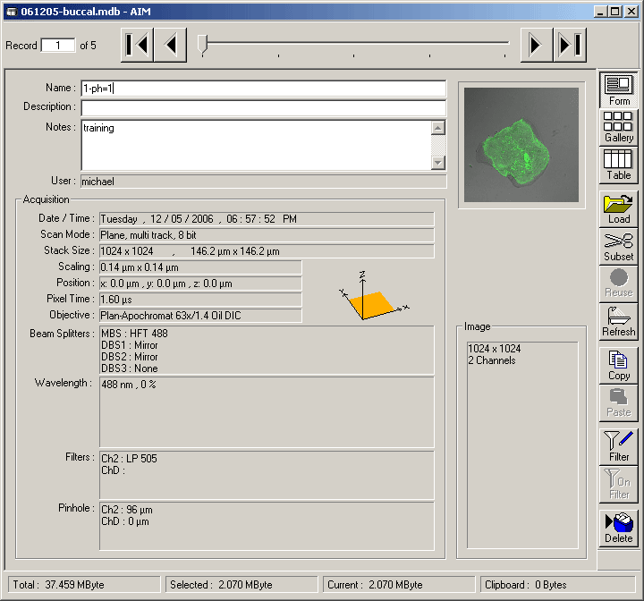
The standard method to save scanned images on the Zeiss 510 confocal microscope is the do a "save" or "save as" which prompts for a name to save the scan as, and also prompts for a named data base to save the named scan to. The database consists of a named directory, which contains .lsm files (multipaged images), one corresponding to each scan, and an .mdb (an Access database file containing scanning parameters), and transiently a .ldb file (database lock file) when the database is being used. Although it is possible to save a scanned image directly as a tiff format file, it is better to save the scan to an image database since scanning parameters, such as object magnification, zoom factor, laser powers, PMT gains, filter settings are stored into the database for each scan. The database information allows tasks such as adding scale calibrations to be done later and the scanned images to be viewed in chronological order with multiple channels overlaid or z-series or time lapse scans to be viewed using the free ZIB v 4.2.0.121 which can be installed on Windows PCs. Also several 3rd party programs (e.g. Volocity, Image Surfer, ImageJ) can be used to view images and effectively read the image database parameters.
Cautions:
Renaming: The files of the image database should not be renamed or modified outside of the Zeiss LSM Image Browser or AIM confocal programs. File names roughly follow the names assigned to the scans while imaging, but with some differences. Reserved characters in the Windows file system can be used in the scan name, but not the actual file name in the database directory. Also more than one scan may have the same name in the database, but the corresponding file names in the directory are necessarily made to be different. However the directory name may be changed with no consequences.
Exporting: Database images may be exported typically as tiff images and viewed in Photoshop, loaded into Power Point, used for publication e-submission, etc. Exporting has some scaling considerations which are described below. Macros for bulk exportation of images are available on the Zeiss 510 confocal computer (Hertz) and the offline AIM LSM software on the image processing workstation called Snell.
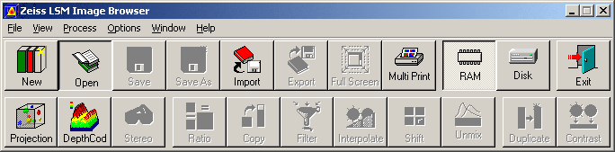
The Zeiss LSM Image Browser version 4
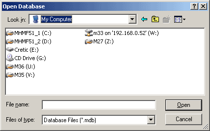
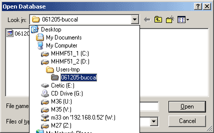
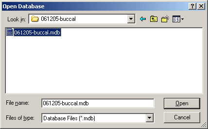
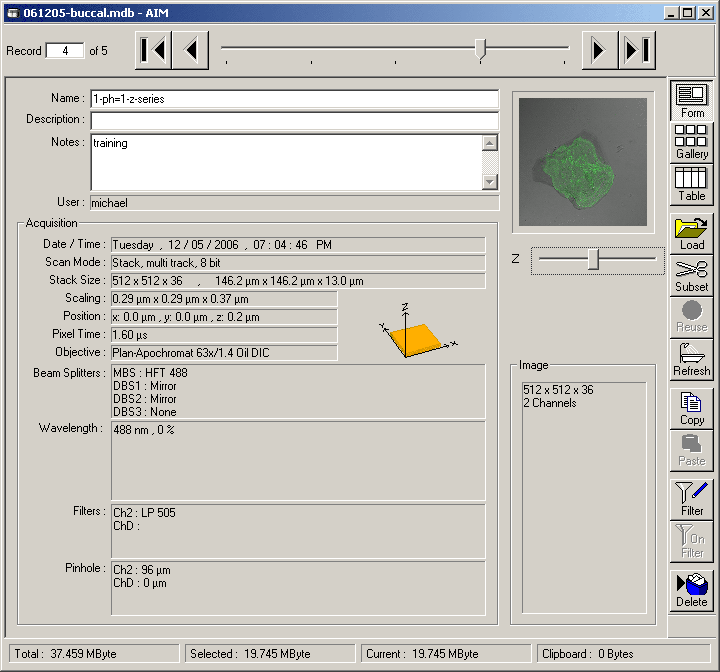
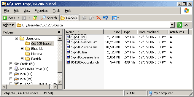
The sample Zeiss image database used on this page can be downloaded in compressed from here 061205-buccal.mdb.zip. Winzip can be used to uncompress the download file.
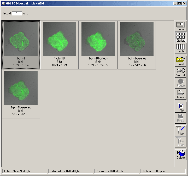
Form view Gallery view Use black left or right pointing triangle to page through database scans Double left click on a thumbnail view to open the scan
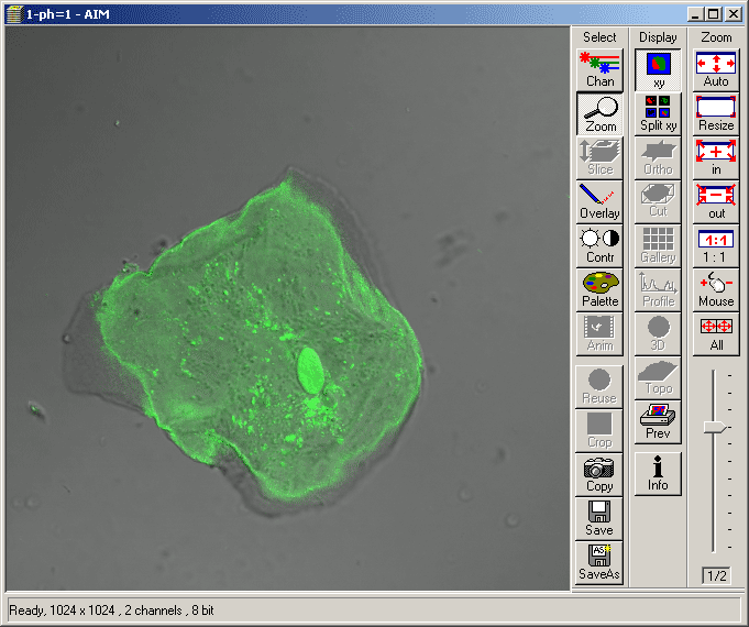
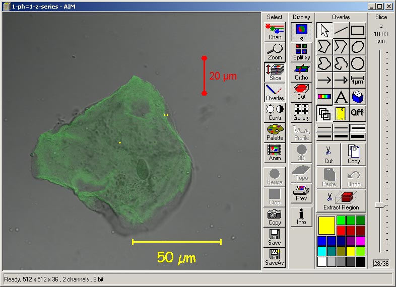
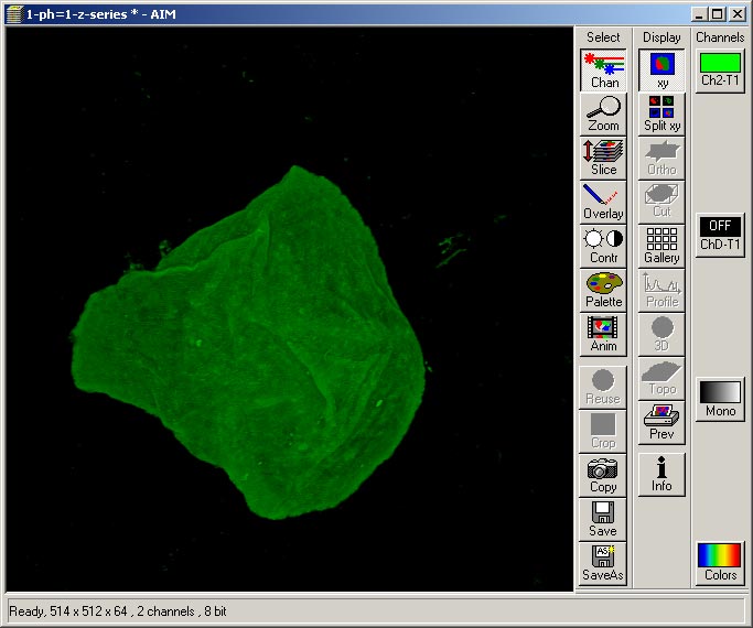
Channels may be removed from the overlay by turning off the undesired channel(s). e.g. Depress Chan, press ChD-T1 and choose Off
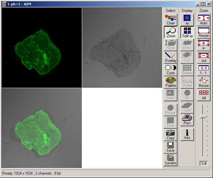
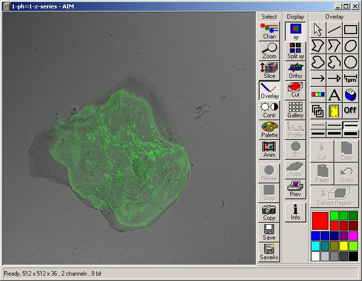
The 2 channel image below has a gray scale palette (with red for overload and blue for under load) for both channels
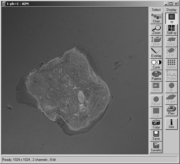
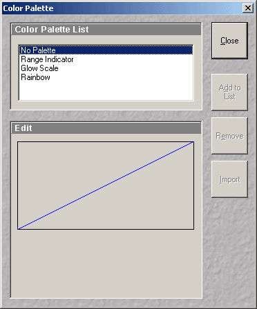
Choosing the "No Palette" option from the Color Pallet List
Now the fluorescence channel(s) is(are) colored and the DIC/transmitted light channel is gray
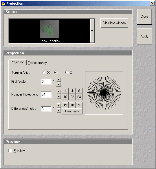
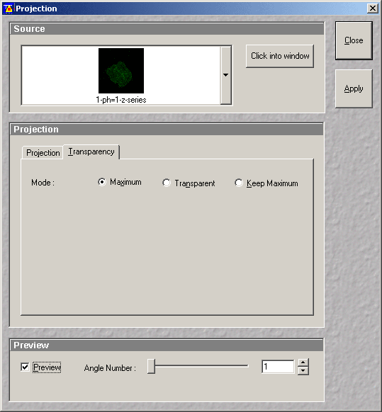
Choose Projection view - for example choose first angle = 0, number of projections = 64, difference angle click Panorama
Check Preview, ensure Source is the correct image stack, and press Apply
![]()

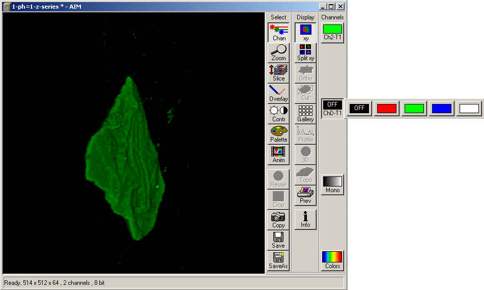
This view is the render of the fluorescence channel only
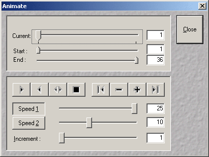
Hit Animate to view a dynamic rotation.
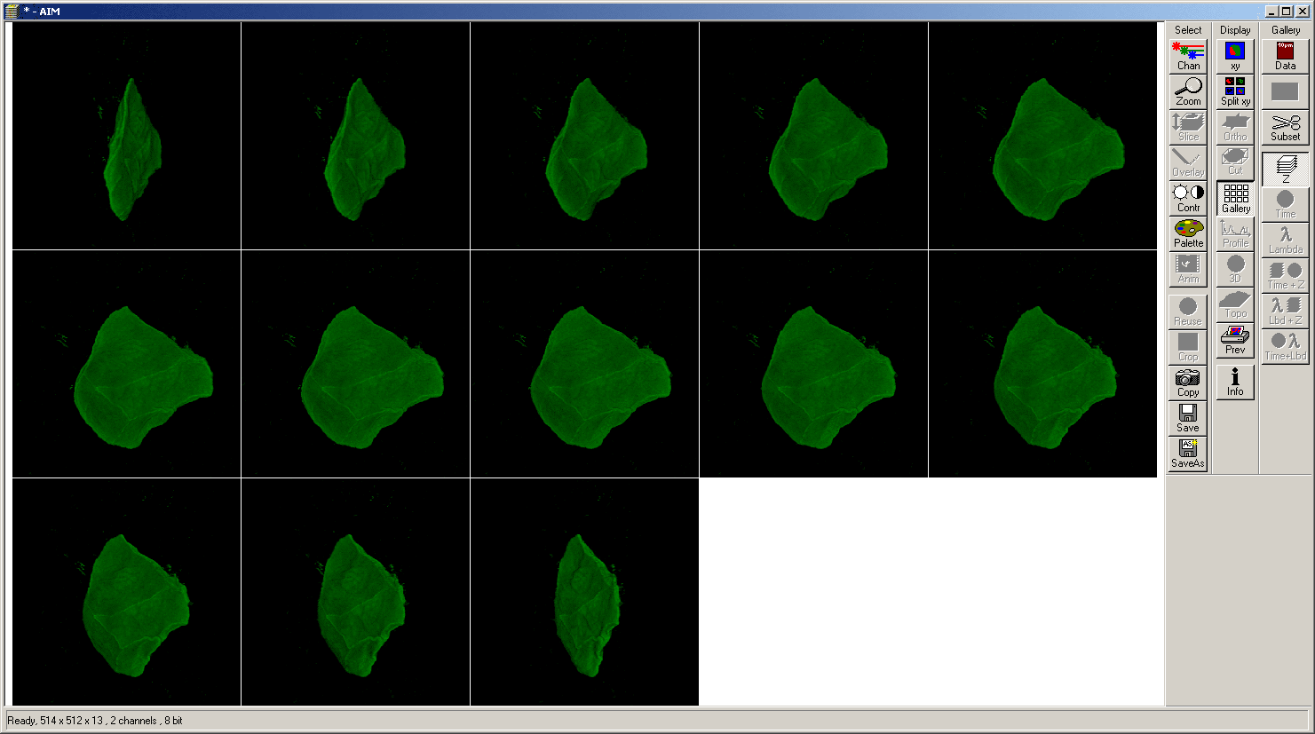
Also postage stamp view of each rendered view can be displayed by hitting the Gallery button
It is possible to see a 3D view in the above gallery by crossing ones eyes. Conditions which make a stereopsis view difficult or impossible include astigmatism, amblyopia, hyperopia, presbyopia, strabismus, less than two functional eyes.
|
|
|
Copyright 2001-2015 Dr. M. Chua, School of Medicine, University of North Carolina, Chapel Hill, NC 27599 |
| Go Back | Booking Resources |
Questions/comments/problems: Michael Chua |
|
|
Last Updated: 2014-07-24 |