Notices
|
|
Notices:
Visiopharm has been updated to version 2023.09 x64 and has
been moved to a faster computer. Each user needs to
reconnection to shared file/server resources.
The Olympus VS200 slide scanner software has been upgraded to
version 3.4.1 on May 19, 2023Please update your OlyVIA
viewer software to version 4.1.1
Please use a recent version of FIJI
Cisco AnyConnect version
4.10.06090, or later, is now required to connect to
the campus VPN. This version requires Windows 8 or later or
Mac OS 10.14 or later to run. |
|
|
|
|
|
|
|
|
|
|
|
|
|
|
| |
|
|
 |
- Marsico Hall 7th floor
- Thurston Bowles room 6024
|
|
|
- Michael 919-912-9380 voice/text
|
|
|
|
|
|
| Confocal First Light - mixed
pollen grains |
.jpg) |
|
5 Channels -
CY2
CY3
CY3.5
CY5
CY7
|
| Excitation - White Light Laser 20x 0.75NA |
|
| Magenta |
Darkfield |
| Yellow |
Confocal reflection |
| Blue |
DAPI |
| Green |
Fluorescence |
| 40x 1.25 NA adjusted to ~0.7 |
 |
|
 |
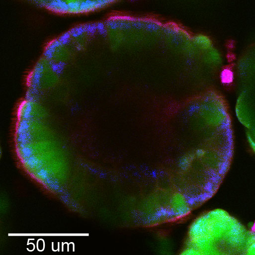
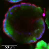
Spheroid
Confocal scan 40x NA 0.6 |
|
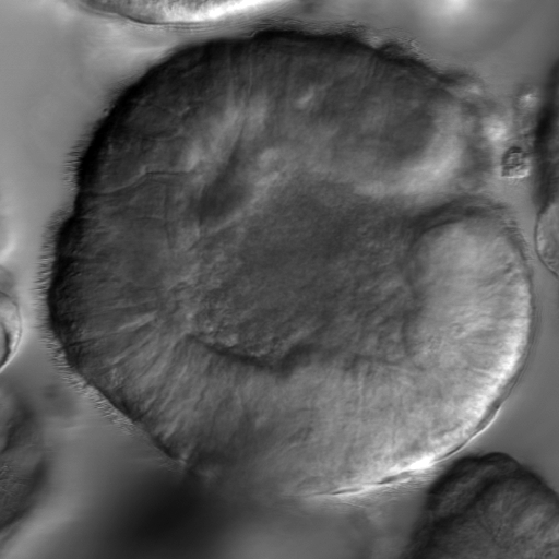
Spheroid
DIC |
|