Michael Hooker Microscopy Facility (MHMF.ORG)
![]()
|
|
Michael Hooker Microscopy Facility (MHMF.ORG) |
|
Image Gallery (click on window to enlarge or play movies)
|
|
|
|
Thin transverse section of a Neuro Muscular Junction. Best wide field view of a z-series |
Same section as left deconvolved on a DeltaVisonRT imaging system |
| Red label is Alpha-bungarotoxin TexasRed. Green label is con-A FITC. Click on image to view full field of view. Sample prepared by Dr. Neal Kramarcy. Image scanned and deconvolved by M. Chua. | |
 |
|
Well differentiated airway epithelial cells cultured on a membrane. NaK-ATPase (green) is on the basolateral membrane. Nuclei are labeled with Propidium Iodide (red). Cilia (gray) above apical membrane (light gray) are imaged using DIC. Tissue prepared and imaged on a confocal microscope by Dr. A. Livraghi, Cystic Fibrosis Center, UNC. |
|
|
|
||||||||
|
|
|
|||||||
|
Arabidopsis guard cell labeled with GFP targeted to the membrane. Images scanned on a Zeiss 510 meta confocal microscope. Sample provided by Shouling Xu. |
|||||||||
|
|
 |
|
| (click on image to see a larger animation) | ||
| A live root tip of Arabidopsis. Plasma membrane labeling with FM 1-43 simultaneously scanned with DIC using a Zeiss 510 confocal microscope. | Arabidopsis guard cell labeled with GFP targeted to the membrane. Rendered with Volocity 2.6.1. | Rendered with T3D |
| Please note that these images are not intended as a comparison of Volocity and T3D. Several factors including transparency and threshold are not equivalent. | ||
| Below: 3 color z-stack antibody localization in epithelial cell tissue culture, using Zeiss Meta Multitracking, with minimal intensity lasers. Image generated by Alan S Fanning, Ph.D. Cell and Molecular Physiology and Tim Oliver | |
 |
Tight Junctions are Disrupted in Epithelial Cells Expressing an Altered ZO-1 Transgene. A Madine-Darby Canine Kidney (MDCK) cell line expressing a myc-tagged ZO-1 transgene was fixed in Ethanol/Acetone and stained with antisera against the c-myc epitope tag (Cy2-green), the transmembrane protein occludin (Cy3-red), and an antiserum specific for the canine ZO-1 (Cy5-blue). The cells were imaged with a 100X PlanApo lens on the Zeiss 510 LSC Microscope using the Meta detector. Note that the transgene (red) forms large ectopic structures distinct from the normal circumferential distribution of the endogenous ZO-1 polypeptide (blue), and that the transmembrane protein occludin is also recruited into these extopic structures (green). |
| Dr. Alan Fanning | |
 |
Maximum projection of a z-series scan through a mouse embryo. Scanned on a Leica SP2 confocal microscope |
|
Mouse embryo |
|
|
Z-series scan acquired using a Zeiss 510 confocal microscope. Render using Volocity 2 made by Dr. Michael Chua Quick Time version of movie (855 KB) |
|
Arabidopsis root hair |
 |
|
|
|
| Lung airway mucus Dr. Hiro Matsui |
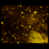
Fluorescent beads in fluorescent mucus |
Mucus above airway cells Dr. Ray Pickles |
Airway cilia. DIC sequence acquired at 3.9 fps using a laser scanning confocal microscope, Dr. P. Sears & A. Rossi |
 |
 |
| Triple Antibody labeled sensory cortex, Dr. Sejin Huang |
Coronal section of spinal cord |
|
|
|
Ice storm of December 2002 |
|
|
|
Copyright 2001-2015 Dr. M. Chua, School of Medicine, University of North Carolina, Chapel Hill, NC 27599 |
| Go Back | Booking Resources |
Questions/comments/problems: Michael Chua |
|
counter |
Last Updated: 2014-07-24 |