Michael Hooker Microscopy Facility (MHMF.ORG)
![]()
|
|
Michael Hooker Microscopy Facility (MHMF.ORG) |
|
Nikon Microphot SA
Fluorescence Filters
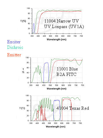 This figure
shows the transmission/wavelength curves for the fluorescence filters that
comprise the three epifluorescent cubes in the rotating carousel above the
objective nosepiece. Each cube contains a set of 3 separate glass filter
elements, the exciter, the dichroic and the emitter.
This figure
shows the transmission/wavelength curves for the fluorescence filters that
comprise the three epifluorescent cubes in the rotating carousel above the
objective nosepiece. Each cube contains a set of 3 separate glass filter
elements, the exciter, the dichroic and the emitter.
The shorter wavelength band pass filters correspond to the exciter (blue lines) and longer wavelength emitter (barrier) filters (red lines) occupy mutually excluding regions of the spectrum, which are separated by the dichroic reflector, the rising green lines.
Naming of filter sets is often confusing, different microscope manufacturers having different labeling conventions for the same filter sets. Also, limited space on the scope itself makes it difficult to identify a cube. So, for example, on the front plate of the cube carousel they are labeled G, UV and B2, where G stands for Green, the color of the excitation light for the cube intended for detecting Rhodamine, UV is the exciting light of the cube for viewing DAPI and B2 stands for Blue, the exciting light for FITC (also GFP, bodipy etc.). Confusing, right? Just remember that the exciting light is the light one sees spilling onto the stage. Note the color which corresponds to the selected filter set. Exciting light is far in excess of the emitted light when viewing the stage with the naked eye.
In current production model microscopes, most vendors now use filters made by Chroma or Omega.
More information about choice of filter sets for particular
biological fluorophores is available on the
Chroma and Omega websites.
|
|
|
Copyright 2001-2015 Dr. M. Chua, School of Medicine, University of North Carolina, Chapel Hill, NC 27599 |
| Go Back | Booking Resources |
Questions/comments/problems: Michael Chua |
|
|
Last Updated: 2014-07-24 |