|
The Michael Hooker Microscopy Facility
is a research light microscopy facility providing advanced digital light microscopy, image processing
and analysis resources to users from the UNC
Chapel Hill campus.
We offer instrumentation and instruction to
enable users to acquire, process and analyze images from a wide variety
of samples, which they provide. |
- DIC/Nomarski Phase contrast/transmitted
- Fluorescence / DIC combined
- Confocal - fluorescence/reflection
- 3-D & reconstruction
- FRAP - FRET
- Live cell imaging (time lapse, 4-D)
- Particle tracking
- Laser Micro-Dissection
- Ratio imaging (Ca++, pH)
- Combined fluorescence & DIC
- and more......
|
| |
|
 |
|
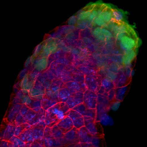
Mouse embryo, Dr. Jaime Rivera |
|
|
|
 |
|
Airway Epithelium, Dr. S. Kreda |
|
| Location |
- Thurston Bowles building, room 6129,
School of Medicine - at the corner of Manning Drive and
South Columbia
|
| Resources available: |
- Zeiss 510 Meta & Leica SP2 aobs
Confocals
- Spinning disk confocal (Perkin Elmer
UltraviewLCS)
- Laser micro-dissection system (Leica AS-LMD)
- Inverted and upright fluorescence/transmitted /Nomarski
microscopes with b/w and color digital cameras
- Leica MZ16FA Fluorescence dissecting
microscope (motorized)
- Image Processing Workstations - 2D, 3D & 4D
software - e.g. Volocity C-Imaging Metamorph
- Heated stages & controlled atmosphere stage
- Humidified incubator for live sample storage
|
| More information
contact: |
- 6007 Thurston Bowles
- 843-3268
- http://microscopy.unc.edu
|
|